
How do x-rays work?
Modern x-ray inspection systems are highly advanced and effective and routinely used in a range of industries for quality control purposes.
Most people, familiar with medical diagnostics and security x-ray systems, are unaware of their significant presence in the food, pharmaceutical and electronics industries where they are quietly working away 24/7 to ensure product quality.
But how do x-ray systems actually produce an image and allow us to see foreign bodies and other product defects?
What are x-rays?
X-rays are electromagnetic radiation, part of a single continuum known as the electromagnetic spectrum, this is arranged according to frequency and wavelength, and runs from radio waves at one end (which have a long wavelength) to gamma rays at the other (which have a short wavelength).
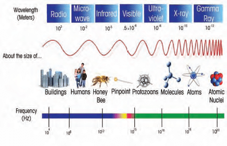
The short wavelength of x-rays enables them to pass through materials that are opaque to visible light, however they do not pass through all materials with the same ease. The transparency of a material to x-rays is broadly related to its density – the denser the material, the fewer x-rays that pass through. Hidden contaminants and other dense internal structures, including foreign bodies like glass, bone and metal, show up because they absorb more x-rays than the surrounding product.
The principles of x-ray inspection
In simple terms, an x-ray system uses an x-ray generator to project a beam of low- energy x-rays onto a linear x-ray sensor or an x-ray imaging intensifier.
The system is typically configured with the x-ray generator above the conveyor, which is used to transport the product through the system and the x-ray sensor positioned under the conveyor which collects and converts the x-ray “shadow graph” signals into a visible image.
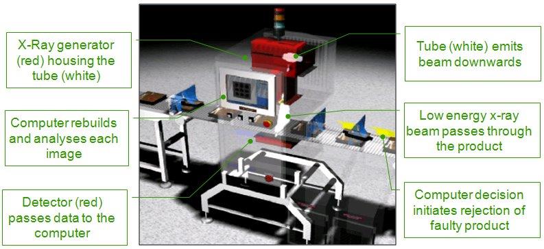
The x-ray shadow graph is generated as a result of the different attenuating properties to the product under inspection. For instance a fragment of glass in a pack of cheese has a higher attenuation (adsorption) than the cheese in which it is embedded therefore as that section of the pack is inspected the area of glass generates a “shadow” or lower beam intensity that the detector can record and display.

It is by this very simple principle that all x-ray systems operate.
In food products the contaminant generally has higher absorption and therefore generally displays as darker than the background image, yet it is still possible to detect material that has a lower absorption than the background. This allows the detection of voiding in filled packs and the detection of missing components in multiple unit packs.
The same shadow attenuation principles apply for the inspection of pharmaceuticals, broken tablets, missing leaflets etc. and across a whole range of industries and quality applications that are beyond the scope of this brief introduction.
X-ray image generating detectors
There are numerous types of x-ray image generating devices but broadly they fall into two categories:
Line array sensors
A line sensor, also often referred to as a linear array, photo-diode array or line scan detector. As its name implies it is a line of x-ray sensitive detectors which is positioned under the conveyor and across it at 90 deg to the direction of travel of the product. It relies on the motion of the conveyor to generate the image in the travel direction and is the most commonly used method in automated systems.
The advantage of these detectors is that they produce a high contrast low, noise image and operate over a wide energy range. The disadvantage is that as they are made up of a single line of individual sensing elements the spacial resolution becomes a function of the sensor element size.
In security applications such as baggage screening the typical size is 1.6 x1.6mm, in food and industrial inspection, typically 0.8 x0.8mm , 0.4 x 0.4mm and in some cases 0.2 x 0.2mm. There are higher resolution sensors than this but it is unusual to need to deploy them in most applications. However AIS do use much higher resolution sensors for recovery applications.
Area sensors
These can either be image intensifiers of flat panel arrays. In fully automated systems these sensors are unlikely to outperform the line sensors. They were developed for and are generally used in the medical sector – for heart procedures and as a replacement for x-ray film.
The main advantage they have is that a full frame image is generated which is not dependent of movement of a conveyor; in fact movement can be detrimental to the image quality if it is not properly controlled. The other advantage of the image intensifier is that it is a very high gain device with high spatial resolution but at the cost of significant signal noise.
In conclusion both techniques have their advantages and at AIS you will always have access to the most appropriate x-ray inspection system for your requirement.
How safe is x-ray inspection of food?
Food that has undergone x-ray inspection remains safe to eat and is indistinguishable in every respect from food that hasn’t been x-rayed. Leading food manufacturers across the world rely on x-ray technology for detection of physical contaminants and for quality control.
Discover more about the facts:
- Scientific evidence (by WHO) states that food radiation levels up to 10,000 Gy does not affect food safety or nutritional value
- EU Directive 1999/2/EC permits x-ray inspection which delivers a dose of less than 0.5 Gy to food products
- The typical dose of radiation for an x-ray linescan system, such as at AIS, is less than 0.2 milli-Gy (one ten millionth of those used in the WHO study)
- Organic food retains its organic status after x-ray inspection
- The irradiation dose that would be required to destroy bacteria in food is much higher than the x-ray inspection dose